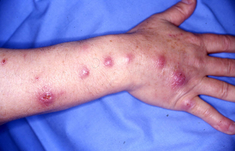
All home hobbyists should be aware that Mycobacterium marinum (the bacteria responsible for most cases of “fish TB”) can infect humans. It typically presents in humans as a very slowly developing (weeks or months) lesion which looks like a big pimple, abscess or carbuncle.
Typical Disease Progression
This is the typical disease progression seen with mycobacterium marinum in a human hobbyist:
Stage #1 First off there will be a red and inflamed hard nodule or even an open abscess on a finger or the back of a hand. It might or might not be painful. The disease may have entered the hand through a cut or a hang nail typically at least three weeks prior. If the initial entry point hasn’t yet healed it probably isn’t mycobacteriosis. The one predictable constant about mycobacteriosis is that it moves VERY slowly.
Stage #2 The hobbyist will go to the doctor. And the doctor will look at it and prescribe an antibiotic. This is exactly what the doctor should do as the vast majority of these abscesses will simply be “normal” bacterial infections. And most of the time the abscess will go away in a week or two and all is well.
Stage #3 Occasionally the antibiotics won’t work and the patient goes back to the doctor. The doctor, again quite correctly, will switch antibiotics as the vast majority of the time it was just a “normal” bacteria which didn’t respond to the first antibiotic (happens all the time, not a big deal, even the best broad spectrum antibiotics only work against about 70% of the pathogenic bacteria). The doctor might order a round of “cultures” taken to see what bacteria they are dealing with. Sometimes even a third round of different antibiotics will be ordered. All will be quite normal and no cause for alarm. The important thing is that one needs to be persistent and keep going back to the doctor till it is cleared up. Easily 90% of the these infections are just a “normal” pathogenic bacteria and will go away with antibiotics.

Stage #4 If a patient has gone through two rounds of antibiotics and the abscesses are still there, then is the time to start mentioning “aquariums” and “mycobacterium marinum” over and over again. If small red spots start appearing every few inches in a line up the arm, a patient needs to get more persistent. It MAY progress up the arm as shown above, typically VERY slowly, like over a span of six months to several years. And sometimes these spots will break open and ooze. These MAY be mycobacterium tubercles or they MAY just be due to a “normal bacteria” which isn’t responding to antibiotics or it MAY be a fungus.
Stage #5 Typically three to four months after the initial doctor’s visit, the disease MAY well be confirmed as mycobacterium by six to ten week long specialized tests. Treatment with an antibiotic cocktail should then begin. Treatment will typically last six months and there may well be some unpleasant side effects.
The important thing here is to not give up. Be persistent but do not panic or get impatient with the doctors. This is a very slow moving disease. If one has mycobacteriosis, it typically will be at least three months before any doctor can tell you “Yes, you have mycobacterium marinum and here is what we will do to treat it“. That is just the way it is. There is no way to speed up the process.
If one gets frustrated with the process and quits going to the doctors, the infection can slowly move into the inside of the body and start creating all sorts of havoc. The key here is persistence without panic.

Mycobacterium in Humans in More Depth
From the paper: “Treatment of Mycobacterium marinum Cutaneous Infections”, Rallis et. al 2007:
“M. marinum infections may have several clinical presentations, but none of these are considered pathognomonic. It is usually presents in immunocompetent patients as a solitary, red to violaceous plaque or nodule with an overlying crust or verrucous surface that may ulcerate. Inflammatory nodules or abscesses can develop in severely immunosuppressed patient, usually in a sporotrichotic type of distribution. This ‘sporotrichoid’ disease begins with distal inoculation and may lead to the development of nodular lymphangitis. Over a period of months, localized cutaneous disease can spread to deeper soft tissues, causing tenosynovitis, arthritis, bursitis and/or osteomyelitis of the underlying bone; it can also be life-threatening, and lesions may or may not be painful.“
Mycobacterium marinum (the bacteria responsible for most cases of “fish TB”) is understudied and probably very under-reported. The best study appears to be that of the Mayo Clinic. In the 21 years between 1998 and 2018 they had forty six cases of Mycobacterium marinum. “Fishing for a Diagnosis, the Impact of Delayed Diagnosis on the Course of Mycobacterium marinum Infection: 21 Years of Experience at a Tertiary Care Hospital”, Castillo et. al., 2020
“Mycobacterium marinum or “fish tank granuloma” is a pathogenic, nontuberculous mycobacterium (NTM) that has been associated with skin, soft tissue, joint, bone, and disseminated infections. It is an endemic fish pathogen widely distributed in aquatic environments such as fish tanks, swimming pools, and natural bodies of water. Despite increasing numbers of cases reported in recent years, the diagnosis of M. marinum is often missed or delayed.
Forty-six cases of culture-confirmed infection with M. marinum were included. Twelve cases (26%) presented with a complicated M. marinum infection. The median time to diagnosis (IQR) was 3.6 (2.3–6.1) months. Only 15 cases (33%) had documented water exposure during the initial evaluation, compared with 33 cases (73%) after microbiology diagnosis was confirmed.”
So this is roughly two per year at one of the thousands of hospitals in the USA. This would indicate this infection is MUCH more prevalent than many give it credit for, on the order of several thousand cases per year in the USA. Note that, even at the advanced hospital that Mayo is, the mean time to getting a diagnosis was 3.6 months. Also note that this study mentioned that one of the problems is that no one asks a patient if they have aquariums when they go to a doctor. So no one makes the link between sores on the hand and arm and Mycobacterium marinum. Duke University Medical Center has a very similar study which came to the same conclusions.
There are 7.2 million households with fish tanks in the USA (13 million aquariums). 2,000/7,200,000 =278 chances in a million that one will get mycobacteriosis from your aquarium in any given year. If you have kept aquariums for 54 years, like I have, that becomes a 1.5% chance one will have contracted this disease. This is much higher than I initially thought. Interesting!

Infections with M. marinum are serious, requiring an average of six months of treatment with several antibiotics. Sometimes the disease penetrates deeply into the tissues and requires surgical intervention and loss of limb function. And rarely people die. So all hobbyists should be aware of it and should mention it to any doctor if they have an unusual slow moving skin infection that looks anything like the above tubercles.
This bacteria does not show up in any normal test like a blood test or a culture. And it is so rare most doctors have never even heard of it, let alone treated a case. This creates a bit of a problem. One will have to be patient if one has tubercles like the above photo. The way M. marinum is diagnosed is by throwing several different antibiotics at it over the course of several months and seeing if one of the antibiotics works to clear it up. i.e.:
“Key diagnostic elements for M. marinum infections are a high index of suspicion raised by negative bacterial tissue cultures, poor response to conventional antibiotic treatments and a history of aquatic exposure”
If several different antibiotics fail to clear up the infection, more advanced tests will be done. These advanced tests take six to ten WEEKS to complete. And even then these tests give false negatives (i.e. the tests say you don’t have the M. marinum when you do have it) 30% of the time. And there are other slow moving diseases such as some fungal diseases which CAN, in some cases, give very similar symptoms. These diseases include:
- Sporotrichosis (also known as “rose gardener’s disease”) is caused by a fungus which lives throughout the world in soil and on plant matter such as sphagnum moss, rose bushes, and hay
- Blastomycosis is caused by a fungus found in moist woods in Northern Climates
- Coccidioidomycosis is caused by a fungus found in Southwest USA
- Histoplasmosis caused by a fungus in soil that contains large amounts of bird or bat droppings in the central and eastern states in the USA
- Leishmaniasis is a parasitic disease common in the tropics. If one has been in say Costa Rica within the past two years this is a possibility.
ALL FIVE of these diseases CAN have the same skin lesion symptoms as mycobacteriosis. All five are resistant to antibiotics. All five are very difficult to test for. All five are very difficult to treat. And the fungal diseases can be “idiopathic”. “Idiopathic” means that one can get a disease like sporotrichosis without ever having been near soil, sphagnum moss, rose bushes, or hay.
So that gives you some idea of what doctors face when presented with such slow moving abscesses. If you are both a gardener and have aquariums things can get obviously rather difficult. And if one is immunocompromised, such as with HIV or diabetes, things get even more complicated.

One just has to be persistent in obtaining resolution. And keep emphasizing to all the doctors that you have an aquarium. Do not give up. If it begins to move up the arm and the doctors won’t listen, go to a teaching hospital. Teaching hospitals are the best place to go for something unusual like this.
Some people in the industry wear long sleeved rubber gloves when working with tank water. Personally I think it is quite sufficient for a worker or a hobbyist to simply know about the disease and to be prepared to tell a doctor if they get an unusual abscess which won’t heal.
Fish TB in More Depth
For those interested in going into more depth on fish TB, clicking on the following links will be useful:
.
.
Aquarium Science Website
The chapters shown below or on the right side in maroon lead to close to 400 articles on all aspects of keeping a freshwater aquarium. These articles have NO links to profit making sites and are thus unbiased in their recommendations, unlike all the for-profit sites you will find with Google. Bookmark and browse!

